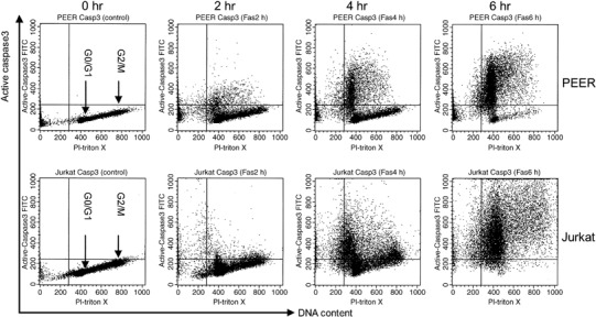
Figure 1.

DNA content and active caspase‐3 double staining analysis of leukemia cells treated with anti‐Fas antibody. Cells (PEER and Jurkat) were pretreated with anti‐Fas antibody (50 ng/mL) for 0, 2, 4 and 6 h. After incubation, cells were harvested and stained with fluorescein isothiocyanate‐conjugated antiactive caspase‐3 antibody and propidium iodide (PI)–Triton X for DNA content, then analyzed by flow cytometry. Similar results were obtained in three repeated experiments.
