
Figure 2.
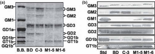
Ganglioside composition as determined by thin layer chromatography (TLC) and TLC‐immunostaining. (a) Gangliosides were examined in TLC using chloroform/methanol: 0.22% CaCl2 (55:45:10). Resorcinol spray was performed for detection of bands. (b) TLC‐immunostaining of gangliosides extracted from four cell lines was performed. After being developed on high performance TLC (HPTLC) aluminium sheets, gangliosides were blotted and then stained with anti‐ganglioside antibodies. Membranes were incubated with first antibodies for 1 h and with second antibodies for 45 min, and then ABC was applied for 30 min at room temperature. Finally, bands were detected with Konica HRP‐1000.