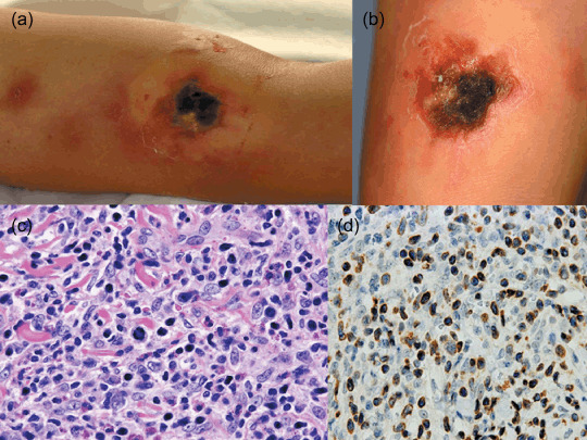
Figure 4.

Cutaneous α/β pleomorphic T‐cell lymphoma. (a) Multiple dark red or brown indurations and an ulcerated induration with black necrotic tissue on the arm. (b) Another ulcerated lesion with hemorrhagic crust and necrotic tissue. (c) Neoplastic cells reveal medium to large pleomorphic nuclei in mitosis and a mixture of reactive lymphocytes, histiocytes, and eosinophils. (d) Neoplastic cells show strong granular staining for perforin.
