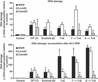
Figure 7.

Phosphorylation levels of γ‐H2Ax as a marker for DNA damage induction in colorectal cancer cells. Western blot analysis was carried out after 72 h incubation with IC50 concentrations. DNA damage accumulation was measured after 72 h drug exposure then recovery for 48 h in drug‐free medium. Values represent means corrected for control phosphorylation levels (set to 1) ± SEM. *Significant differences between treated and control (P < 0.05). E, erlotinib; E → T + E, cells preincubated with E, followed by T + E combination; T → T + E, cells preincubated with trifluorothymidine (TFT), followed by T + E combination; T + E, simultaneous combination of TFT and E.
