
Figure 2.
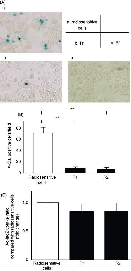
β‐Galactosidase (β‐gal) expression in Ad‐lacZ‐treated radioresistant cells was lower than that in Ad‐lacZ‐treated radiosensitive cells, but there were no differences in adenovirus uptake between radioresistant and radiosensitive pancreatic cells. Cells were infected with Ad‐lacZ at 10 multiplicities of infection (MOI) and β‐gal activity was assessed by X‐gal staining at 48 h after infection. (A) Photomicrographs of X‐gal‐stained radiosensitive or radioresistant cells, ×100. a, CFPAC‐1 parent cells; b, R1 cells; c, R2 cells. (B) Number of β‐gal‐positive cells. Each value represents the mean ± SD of five independent fields. **P < 0.01. (C) Cells were infected with Ad‐lacZ at 10 MOI and DNA was extracted at 24 h after infection. Viral DNA content was quantified by real‐time PCR and defined as the ratio compared with radiosensitive cells. Each value represents the mean ± SD of triplicate measurements.