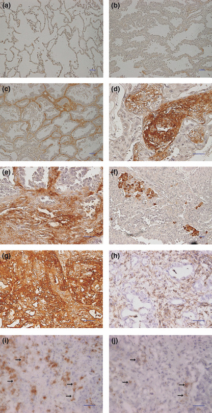
Figure 1.

Nectin‐like molecule‐5 (Necl‐5) expression in primary lung adenocarcinoma tissues. Normal lung tissue and primary pulmonary adenocarcinoma tissues were stained with the anti‐Necl‐5 antibody using the immunoperoxidase technique. (a) Necl‐5 was not expressed in normal lung tissue. (b) Cancer cells were not stained in the bronchioloalveolar carcinomas classified as Noguchi type A. (c) Necl‐5 was expressed only in the stroma in the peripheral area of the bronchioloalveolar carcinoma. (d–f) Cancer cells strongly expressed Necl‐5. (g,h) The serial sections were stained with the anti‐Necl‐5 antibody (g) and the anti‐Vimentin antibody (h) to verify stroma cells expressing Necl‐5. Vimentin was expressed in the stroma cells but not in the cancer cells (h). Comparison of these two slides revealed that Necl‐5 was expressed in both cancer and stroma cells. (i,j); The serial frozen sections were stained with the anti‐Necl‐5 antibody (i) and the anti‐integrin avβ3 antibody (j). Comparison showed that both Necl‐5 and integrin avβ3 were expressed at the same sites (arrow in i and j). It suggested the colocalization of Necl‐5 and integrin avβ3 in the invasive front of lung cancer samples. Scale bar, 50 mm in (a–j).
