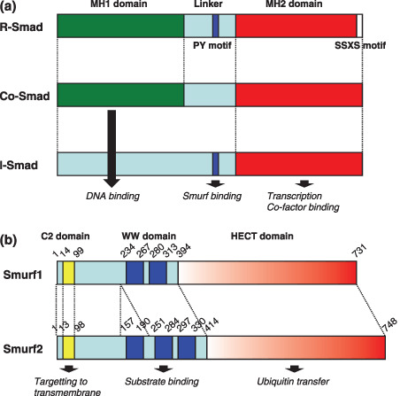
Figure 2.

Schematic diagram displaying the structural organization of Smad proteins and Smad ubiquitin regulatory factor (Smurf) proteins. (a) Diagrammatic representation of the three subfamilies of Smads. Smad proteins consist of two conserved domains, the MH1 and MH2 domains, and the linker region. The PY motif of the linker that is recognized by the homologous to the E6‐accessory protein C‐terminus (HECT)‐type E3 ubiquitin ligases, and receptor‐mediated phosphorylation occurs at the carboxy‐terminal SSXS motif of R‐Smads. (b) Diagrammatic representation of Smurf1 and Smurf2. The C2 domain is responsible for localization of Smurfs to the plasma membrene in a Ca2+‐dependent manner. The WW domain binds substrate proteins containing PY motifs. The HECT domain catalyzes the transfer of ubiquitin to target substrates.
