
Figure 4.
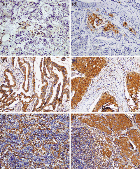
Representative photographs of the immunohistochemical study of S100A11 protein expression in non‐small cell lung cancer specimens (magnification, ×100). (a–c) Adenocarcinoma specimens; (d–f) squamous cell carcinoma specimens. (a) weak staining of lymph node‐negative adenocarcinoma tissues; (b) strong staining of lymph node‐positive adenocarcinoma tissues; (c) strong staining of the matched local positive lymph nodes; (d) weak staining of lymph node‐negative squamous cell carcinoma tissues; (e) strong staining of lymph node‐positive squamous cell carcinoma tissues; and (f) strong staining of the matched local positive lymph nodes.