
Figure 3.
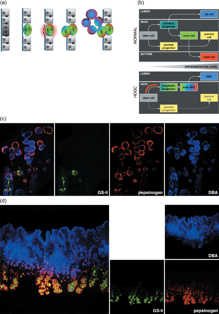
Model of the early development and differentiation of eHDGC. (a) Mis‐orientation of the mitotic spindle and enzymatic digestion of the basement membrane may result in daughter cells dividing out of the epithelial plane and accumulating in the lamina propria. Colors refer to the different gastric lineages as detailed in (b–d). (b) Developmental program of gastric epithelium. The gastric stem cell is thought to reside at the upper isthmus (the upper part of the gastric proliferative zone, the mucous neck region) and give rise to two precursor cells: the mucous neck cell/surface pit cell progenitor and the parietal progenitor. The parietal progenitor produces the lineage of terminally differentiated, acid‐secreting parietal cells found mostly around the neck region of gastric glands. The neck/pit progenitor gives rise to two separate lineages: the terminally differentiated surface pit cells located at the gastric lumen, and the proliferative mucous neck cells at the gastric neck. The neck cells further mature into pepsinogen‐secreting, terminally differentiated chief cells located at the bottom of gastric units (upper). In HDGC, E‐cadherin deficiency causes loss of polarity and, as a consequence, mis‐distribution of gastric cell fate factors (lower). Accordingly, gastric lineages often do not become separated, with neoplastic SRCs expressing markers of up to three distinct cell types including the co‐expression of differentiation markers normally observed at the lumen (pit) or the bottom (chief) of gastric units. Red bar indicates approximate developmental stage at which the second hit can occur. The fate of the parietal lineage during HDGC development is not known. Either the second hit does not affect the gastric stem cell, or E‐cadherin deficiency is not compatible with the survival of the parietal lineage. Notably, diffuse gastric cancer is characterized by a lack of parietal‐type cells. (c) Fluorescence image showing the co‐expression of up to three gastric lineage markers in individual SRCs (GSII/green, mucous neck cells; pepsinogen/red, chief cells; DBA/blue, pit cells).( 11 ) Note simultaneous expression of pit and chief cell markers in the large, luminal SRCs and the peripheral, unpolarized expression of pepsinogen in the SRCs. (d) Birth of intramucosal SRCs from the proliferating region (green) of the gastric antrum. The gastric antrum lacks the typical bottom part of gastric glands enriched with chief cells. Note expression of pepsinogen in the luminal, pit‐type SRCs.