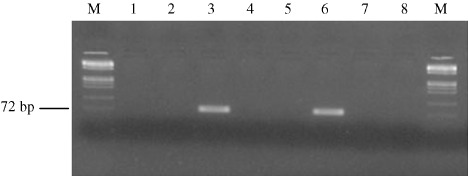
Figure 1.

Detection of API2–mucosa‐associated lymphoid tissue‐1 (MALT1) fusion transcript. The figure shows representative photograph of seminested polymerase chain reaction (PCR) with primers PA4‐R3 for fusion transcript API2 1446–MALT1 814. M, DNA Marker IX (72–1353 bp; Roche Applied‐Science, Indianapolis, IN, USA); lanes 1, 2, 4 and 5, cases that did not show any amplified fragments; lane 3, case of diffuse large B‐cell lymphoma showing a 75‐bp band in the PCR, indicating that API2 1446–MALT1 814 type fusion is present (case USA‐2170 from USA); lane 6, positive control for fusion transcript API2 1446–MALT1 814; lane 7, negative control for RNA extraction; lane 8, negative control for PCR reaction.
