
Figure 1.
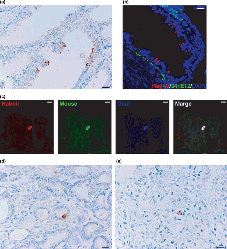
Immunohistochemical analysis of Reg IV expression in clinically localized prostate cancer. (a) Immunostaining of Reg IV in adjacent non‐neoplastic prostate tissue. Several luminal epithelial cells show Reg IV staining. Scale line, 25 µm. (b) Double‐immunostaining of Reg IV (red) and 34βE12 (a marker for basal cells; green). Nuclei are stained with 4′,6‐diamidino‐2‐phenylindole (DAPI; blue). Scale line, 25 µm. (c) Triple‐immunostaining of rabbit polyclonal anti‐Reg IV (red), mouse monoclonal anti‐Reg IV (green), and goat polyclonal anti‐Reg IV (blue). Scale line, 13 µm. (d) Immunostaining of Reg IV in prostate cancer (PCa). Mucin‐like staining of Reg IV is present in goblet cell‐like vesicles of tumor cells. Scale line, 25 µm. (e) Immunostaining of Reg IV in PCa. Perinuclear staining of Reg IV is present in tumor cells. Scale line, 25 µm.