
Figure 2.
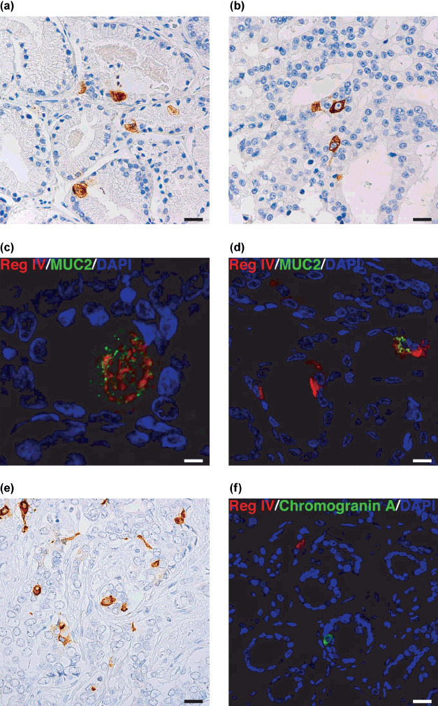
Immunohistochemical analysis of MUC2 and chromogranin A expression in clinically localized prostate cancer (PCa). (a) Immunostaining of MUC2 in PCa. Mucin‐like staining of MUC2 is present in goblet cell‐like vesicles of a tumor cell. Scale line, 25 µm. (b) Immunostaining of MUC2 in PCa. Perinuclear staining of MUC2 is present in a tumor cell. Scale line, 25 µm. (c) Double‐immunostaining of Reg IV (red) and MUC2 (green). Nuclei are stained with 4′,6‐diamidino‐2‐phenylindole (DAPI; blue). Reg IV staining is present with MUC2 in goblet cell‐like vesicles of a tumor cell. Scale line, 13 µm. (d) Double‐immunostaining of Reg IV (red) and MUC2 (green). Nuclei are stained with DAPI (blue). A tumor cell showing perinuclear staining of Reg IV also shows MUC2 staining. Scale line, 25 µm. (e) Immunostaining of chromogranin A in PCa. Scale line, 25 µm. (f) Double‐immunostaining of Reg IV (red) and chromogranin A (green). Nuclei are stained with DAPI (blue). Scale line, 25 µm.