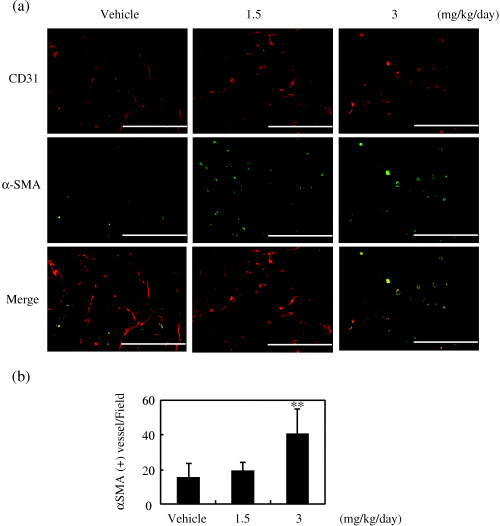
Figure 8.

Effect of RA‐VII on tumor vessel maturation. Tumor sections obtained on day 15 were stained for CD31 and α‐smooth muscle actin (αSMA), as described in Materials and Methods. CD31 was visualized using biotinylated anti‐CD31 and a streptavidin–Cy3 conjugate, and αSMA was visualized with mouse anti‐αSMA and fluorescein‐isothiocyanate‐conjugated goat antimouse antibody. (a) Fluorescent images of CD31 and αSMA. Scale bars = 200 µm. (b) Quantification of αSMA‐associated mature vessels was carried out by counting CD31+/αSMA+ vessels per field. Their numbers were counted and calculated as the mean of five fields per section, and the values expressed as the mean ± SD of four sections. **P < 0.01 versus vehicle.
