
Figure 7.
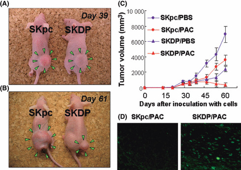
The effect of the overexpression of dipeptidyl peptidase IV (DPPIV) on subcutaneous tumor formation in nude mice with or without treatment with paclitaxel. A total of 2.0 × 10⁶ SKpc and SKDP cells were subcutaneously implanted in the lower flank of nude mice. Subsequent intraperitoneal (i.p.) administration of paclitaxel (20 mg/kg) or PBS was initiated 20 days after tumor inoculation, and repeated twice a week for a maximum of 6 weeks. (A,B) Among paclitaxel‐treated mice, the general appearance of mice 39 (A) and 61 (B) days after inoculation with SKpc (left mouse) and SKDP (right mouse) cells. On Day 39, the tumor size of the SKDP cell‐inoculated group was as large as that of the SKpc cell‐inoculated group. In contrast, on Day 61, the former was smaller than the latter. (C) Tumor growth curves of the four groups: Red closed red circle (SKpc/PAC‐group), SKpc‐inoculated group treated with paclitaxel (n = 7); closed red triangle (SKDP/PAC‐group), SKDP‐inoculated group treated with paclitaxel (n = 7); closed blue circle (SKpc/PBS‐group), SKpc‐inoculated group treated with PBS (n = 7); closed blue triangle (SKDP/PBS‐group), SKDP‐inoculated group treated with PBS (n = 7). On Day 61: P < 0.0001, SKpc/PAC‐group versus SKDP/PAC‐group; P < 0.0001, SKpc/PBS‐group versus SKDP/PBS‐group. Data are presented as the mean ± SD. (D) TUNEL staining was performed for the resected murine subcutaneous tumor on Day 61. Apoptotic cells were identified by positive TUNEL staining (left panel, SKpc/PAC; right panel, SKDP/PAC).