
Figure 1.
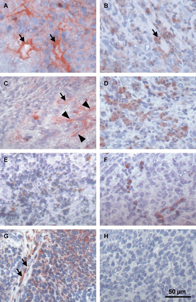
Immunohistochemical analysis of different angiogenic growth factors and the hepatocyte growth factor (HGF) receptor c‐MET in head and neck squamous cell carcinoma (HNSCC). Tumor sections of a hypopharynx carcinoma (#38, Table 1) stained positive for (A) basic fibroblast growth factor (bFGF), (B) vascular endothelial growth factor (VEGF), (C) HGF, (D) platelet derived growth factor (PDGF), (E) granulocyte colony‐stimulating factor (G‐CSF), and (F) granulocyte macrophage colony‐stimulating factor (GM‐CSF). Except for HGF, all growth factors analyzed were found on tumor cells and accumulated around blood vessels (arrows). Arrowheads in (C) point to HGF‐positive stroma cells. (G) Analysis of c‐MET in a larynx carcinoma (#26) reveals expression on endothelial (arrows) and tumor cells. (H) Negative control corresponding to (G).