
Figure 1.
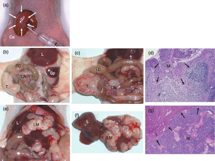
Orthotopic implantation of colorectal cancer (CRC) cells using Matrigel (a), and their macroscopic (b, c, e, f) and histological (d, g) characteristics. (a) The cecal wall bulges slightly after 100 µL injection of Matrigel solution containing 2.0 × 10⁶ CRC cells (white arrows). (b) Eight weeks after implantation of the LoVo cell line. The local tumor growth was recognized at the cecum. The mesenteric LN was not swollen. (c) Eight weeks after implantation of Clone A. The local tumor and a swollen LN were recognized. (d) Pathohistology of LN metastasis shown in (c) (hematoxylin–eosin [HE] staining, 400×). (e) Liver metastasis after implantation of HCT‐15 cells. (f) Liver translocated outside the body. The number of suspicious metastatic nodules was counted and the tissue was prepared with standard HE staining to confirm metastatic cancer cells. (g) Pathohistology of liver metastasis as shown in (f) (HE, 400×). Black arrows in (d) and (g) indicate invasive fronts of cancer cells. Ce, cecum; F, lymphoid follicle; I, ileum; L, liver; LM, liver metastasis; LN, lymph node; M, lymph node or liver metastasis; Sp, spleen; T, orthotopically implanted tumor onto cecal walls; TC, transverse colon.