
Figure 3.
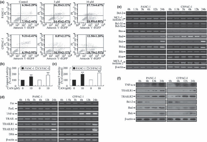
Induction of apoptosis by cantharidin. (a) After cantharidin treatment, the cell populations at both early apoptosis (annexin V+/propidium iodide [PI]−) and late apoptosis (annexin V+/PI+) stages increased significantly in a dose‐dependent manner. *P < 0.05, **P < 0.01, indicate significant differences from the respective control groups. (b,c) Caspase‐8 and caspase‐9 were activated after treatment of cells with cantharidin. *P < 0.05, **P < 0.01, indicate significant differences from the respective control groups. (d) Incubation with 10 μm cantharidin led to time‐dependent alteration of mRNA levels of genes related to extrinsic pathway. The expressions of tumor necrosis factor‐α (TNF‐α), TNF‐related apoptosis inducing receptor 1 (TRAILR1)/death receptor 4 (DR4), and TRAILR2/DR5 increased markedly. (e) Treatment with 10 μm cantharidin altered the expression pattern of genes associated with intrinsic pathway in a time‐dependent manner. The expressions of the anti‐apoptotic gene (Bcl‐2) and pro‐apoptotic genes (Bad, Bak, and Bid) were respectively down‐regulated and up‐regulated. PANC‐1 cells did not express Bik. The expressions of the other investigated genes did not show significant changes. (f) Cantharidin (10 μm) treatment induced time‐dependent up‐regulation of TNF‐α, TRAILR1, TRAILR2, Bad, and Bak, and down‐regulation of Bcl‐2 in both cell lines at the protein level.