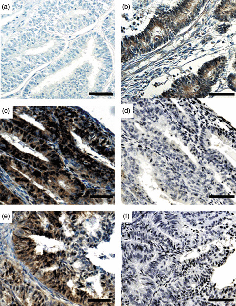
Figure 1.

Representative results of phosphorylated (p)‐AKT immunohistochemistry in endometrial cancer specimens. (a) A sample with negative staining. (b) A sample with moderate expression. (c,e) Cases with strong expression, showing both nuclear and cytoplasmic expression. (d,f) Absorption test using p‐AKT blocking peptide in (d) case C and (f) case E. The staining completely disappeared with the addition of blocking peptide. (Original magnification ×200. Scale bars = 100 µm.)
