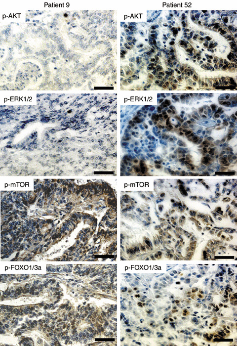
Figure 3.

Concordance of phosphorylated (p)‐AKT and p‐ERK1/2 expression in endometrial cancer specimens. Patient 9 showed negative staining for p‐AKT and p‐ERK1/2, whereas both were positive in patient 52. Staining for p‐mTOR and p‐FOXO1/3a was positive in both patients. Note the concordance of p‐AKT and p‐ERK1/2 expression in each patient. (Original magnification ×200. Scale bars = 50 µm.)
