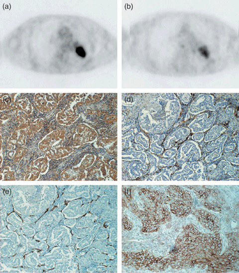
Figure 1.

Transaxial sections of 2‐[18F]‐fluoro‐2‐deoxy‐D‐glucose positron‐emssion tomography (18F‐FDG PET) (a) and L‐[3‐18F]‐α‐methyltyrosine PET (18F‐FMT PET) (b) of a 74‐year‐old man with adenocarcinoma of the left lung (p‐T2N0M0). Tumoral 18F‐FDG and 18F‐FMT uptake is intense with standardized uptake value (SUV) = 11.5 and SUV = 2.65, respectively. Immunostaining for vascular endothelial growth factor (VEGF) (c) reveals that more than 80% of tumor cells show positive reaction for anti‐VEGF antibody. Immunostaining for CD31 (d) and CD34 (e) reveal that many small vessels with positive CD31 or CD34 are seen in the stroma of tumor tissue. The scoring of L‐type amino acid transporter 1 immunostaining was grade 4 and its immunostaining pattern was membranous (f).
