
Figure 2.
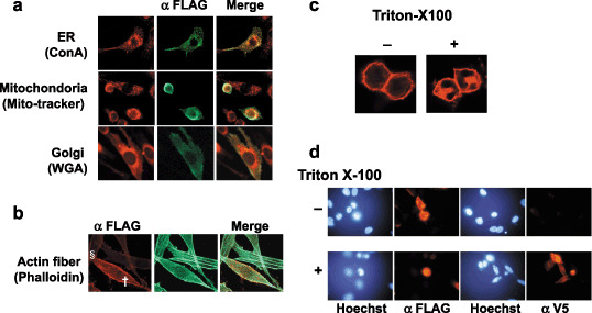
Subcellular localization of SVS‐1 protein expressed in NIH3T3 cells. At 48 h after transfection with pcDNA3‐FSVS‐1, cells were fixed and perneabilized by Triton X‐100 treatment. (a) Fixed cells were costained with anti‐Flag antibody followed by fluorescein isothiocyanate (FITC)‐labeled secondary antibody and probes specifically staining cellular organells, Con A staining endoplasmic reticulum (ER), wheat germ agglutinin (WGA) staining Golgi apparatus, mito‐tracker staining mitochondria. (b) Actin fibers were stained with FITC‐labeled phalloidin, strong (†) and weak (§) expression of SVS‐1 being shown. (c) Fixed cells were washed with phosphate‐buffered saline with or without 0.2% Triton X‐100, stained with anti‐FLAG antibody, and observed with laser confocal microscope. (d) Fixed cells permeabilized or not were stained with anti‐FLAG antibody or anti‐V5 antibody.