
Figure 4.
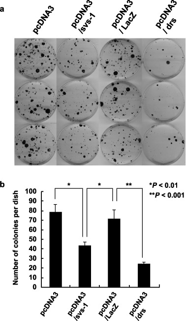
Growth inhibition of Ki3T3 cells by SVS‐1, as assayed by colony formation. (a) Ki3T3 cells were transfected with pcDNA‐Fsvs‐1v5his, pcDNA‐LacZ, pcDNA‐Drs or pcDNA3.1 and plated in medium containing G‐418. Colonies were fixed stained with methylene blue, and counted. The experiments were repeated three times using triplicate dishes. (b) Average numbers of colonies in each group are shown. P‐values are shown.