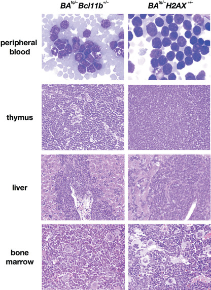
Figure 2.

Representative results of pathological analysis of BA tg/– Bcl11b +/– (left panels) and BA tg/– H2AX +/– (right panels) leukemic mice. Wight‐Giemsa‐stained peripheral blood smears and HE‐stained tissue slices are shown. In both leukemic mice, blast cells proliferated in the peripheral blood (upper panels), caused destruction of the basal structure of the thymus (second panels), and infiltrated around the vessel and in the sinusoids in the liver (third panels). In contrast, bone marrow exhibited myeloid cell hyperplasia with differentiation and proliferation of megakaryocytes (bottom panels).
