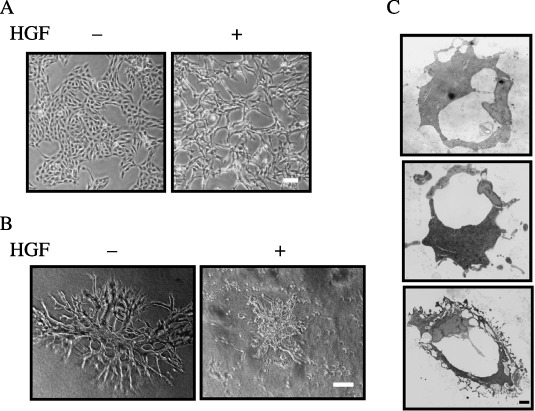
Figure 1.

Morphology of porcine aortic endothelial (PAE) cells in the absence or presence of hepatocyte growth factor (HGF). (A) Morphology of PAE cells in two‐dimensional culture. PAE cells were cultured for 24 h in the absence (–) or presence (+) of HGF (50 ng/mL). The morphology of the cells was analyzed by light microscopy. Scale bar = 10 µm. (B) Morphology of PAE cells cultured in three‐dimensional collagen gel. PAE cells were cultured for 8 days in the absence (–) or presence (+) of HGF (50 ng/mL). The morphology of the cells was analyzed by light microscopy. Scale bar = 10 µm. (C) Electron microscopic analysis of sections of the tubule structure formed by PAE cells in three‐dimensional culture in the absence of HGF. Scale bar = 1 µm.
