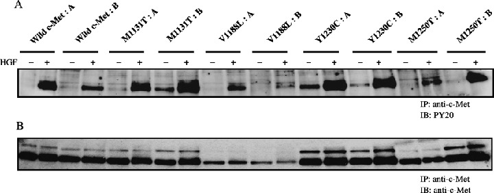
Figure 2.

Immunoblotting analysis of the expression and phosphorylation of c‐Met proteins. Porcine aortic endothelial (PAE) cells expressing c‐Met proteins were treated for 5 min with (+) or without (–) hepatocyte growth factor (HGF) (50 ng/mL). Lysates of the cells were immunoprecipitated (IP) with an anti‐c‐Met antibody and the immunoprecipitates were immunoblotted (IB) with an antiphosphotyrosine antibody (PY20) (A) and with an anti‐c‐Met antibody (B).
