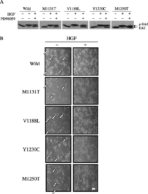
Figure 7.

Effect of PD98059 on the morphology of porcine aortic endothelial (PAE) cells expressing c‐Met mutants in three‐dimensional culture. (A) PAE cells cultured in two‐dimensions were pretreated for 1 h with (+) or without (–) 5 µM PD98059, and treated with (+) or without (–) hepatocyte growth factor (HGF) (50 ng/mL). Lysates of the cells were immunoblotted with an anti‐ERK2 antibody. (B) PAE cells were cultured for 8 days in the absence (–) or presence (+) of HGF with 5 µM PD98059. The morphology of the cells was analyzed by light microscopy. Similar results were obtained in two clones of the wild‐type and mutant c‐Met. The results for one of them are shown. The arrows indicate the short tubule‐like structures. Scale bar = 20 µm.
