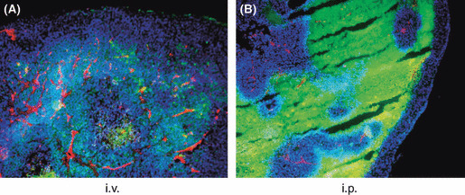
Figure 4.

Accumulation pattern of PTX in peritoneal tumors. Peritoneal tumors were excised at 24 h after i.v. (A) or 48 h after i.p. (B) injection of paclitaxel nanoparticles (PTX‐30W), and vessels were stained red with anti‐PECAM‐1 mAb and nuclei were stained with DAPI as described in the Materials and Methods. Most of the green PTX signal was associated with PECAM‐1‐positive vessels in (A), whereas it was strongly detected in the PECAM‐1‐negative area and the area around PECAM‐1‐positive vessels lacked PTX accumulation in (B).
