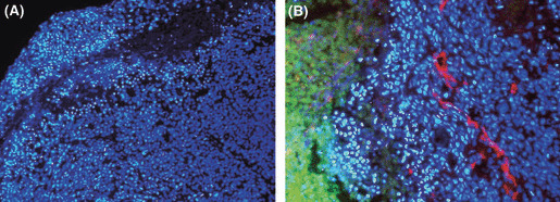
Figure 6.

Apoptosis of tumor cells located in the vicinity of a paclitaxel (PTX)‐accumulated area. Peritoneal tumors were excised at 24 h after i.p. injection of PTX‐30W, and the nuclei were stained in blue with Hoechst 33342 (×200) (A). The nuclear staining was overlayed with the green fluorescent signal of OG‐PTX and counterstaining of tumor vessels with anti‐PECAM‐1 in red (×400) (B). Apoptotic nuclei were condensed with brighter staining with Hoechst 33342.
