
Figure 4.
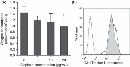
(A) Oxygen consumption of OVCAR‐3 cells after treatment with cisplatin at the indicated doses for 3 h. The results are from three independent replicates. The experiments were repeated three times, and representative results are shown. *P < 0.05 compared with the control. (B) Histogram of mitochondrial membrane potential assessed by MitoTracker 3 h after treatment with 20 μg/mL cisplatin. The y‐axis represents the percentage of maximum cell number counts (Max); the x‐axis represents the mean MitoTracker fluorescence intensity. The dotted line indicates untreated cells. Gray shading indicates untreated cells with the MitoTracker, and the solid line indicates cisplatin (20 μg/mL)‐treated cells with the MitoTracker.