
Figure 6.
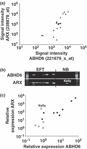
Correlation of expression of α/β hydrolase domain containing 6 (ABHD6) and aristaless (ARX) in Ewing family tumors (EFT). (a) Presented are signal intensities (Affymetrix HG_U133Plus2.0 microarrays) for ABHD6 and ARX in EFT cell lines. (b) Presented are results from conventional RT‐PCR analysis with ABHD6‐ and ARX‐specific primers. ABHD6 signals were only detectable in EFT cell lines, with the exception of the neuroblastoma cell line Kelly, ARX was also detectable only in EFT. β‐Actin (ACTB) was used as a control (not shown). The following cell lines were analyzed: (from left to right) A673, SK‐N‐MC, RD‐ES, SK‐ES, TC‐32, STA‐ET8, TC‐71, TTC‐466 (EFT), SiMa, SH‐SY5Y, IMR5, Kelly, and CHP‐134 (neuroblastoma). (c) Presented are results from quantitative RT‐PCR analysis with ABHD6‐ and ARX‐specific primers. EFT samples are presented as filled circles, neuroblatsoma samples as open circles (same samples as in [b]). For comparative gene expression analysis GAPDH was used as a calibrator. The signal of one neuroblastoma cell line was set as one. The position of the Kelly cell line is indicated.