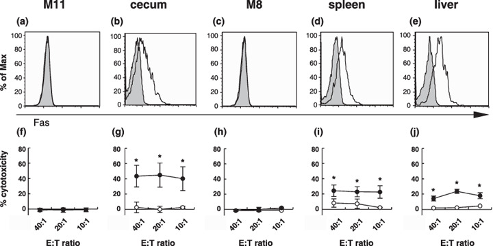
Figure 7.

Tumor cells from the ceca, spleens, and livers expressed Fas and were sensitive to FasL‐induced apoptosis. Fas expression and sensitivity to FasL of (c–e,h–j) SL4‐M8 and (a,b,f,g) SL4‐M11 cells were examined. The cells were grown (a,c,f,h) in vitro and in vivo and were collected from (a,f) ceca, (d,i) spleens, and (e,j) livers. (a–e) The cell surface expression of Fas was examined by anti‐Fas monoclonal antibody binding by flow cytometric analysis. MUC1+ cells that were regarded as in vivo‐growing tumor cells were gated and analyzed. The gray‐filled histograms represent the isotype controls and the open histograms represent anti‐Fas monoclonal antibody binding. (f–j) FasL killing assays were carried out. Target tumor cells were collected from ceca, spleens, and livers by cell sorting using anti‐MUC1 monoclonal antibody (MY.1E12) binding. Parental L5178Y cells (open circle) and mFasL/L5178Y cells (filled circle) were used as effector cells. The percentage of specific 51Cr release was calculated according to the following formula: 51Cr release (%) = 100 × ([cpm experiment – cpm spontaneous release]/[cpm maximum – cpm spontaneous release]). Spontaneous release was obtained from target cells incubated with medium alone and the maximum release was obtained from target cells incubated with 1 M HCl instead of effector cells. Data are represented as the mean ± SD (*P < 0.005). E:T, effector:target.
