
Figure 1.
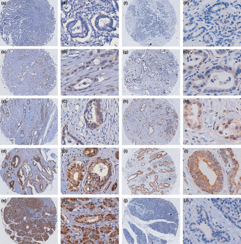
Immunohistochemical evaluation of X chromosome‐linked inhibitor of apoptosis protein (XIAP) expression and XIAP‐associated factor 1 (XAF1) in pancreatic cancers and normal pancreas on tissue microarrays. Invasive pancreatic cancers are shown with negative (a,A), weak (b,B), moderate (c,C), and strong (d,D) XAF1 staining. (e,E) Normal pancreatic tissue showing strong staining of epithelial and acinar cells. (f–i and F–I) Pancreatic cancers showing weak, moderate, and strong XIAP expression. (j,J) Normal pancreatic tissue showing negative XIAP expression. Magnification, (a–j) ×100; (A–J) ×400.