
Figure 3.
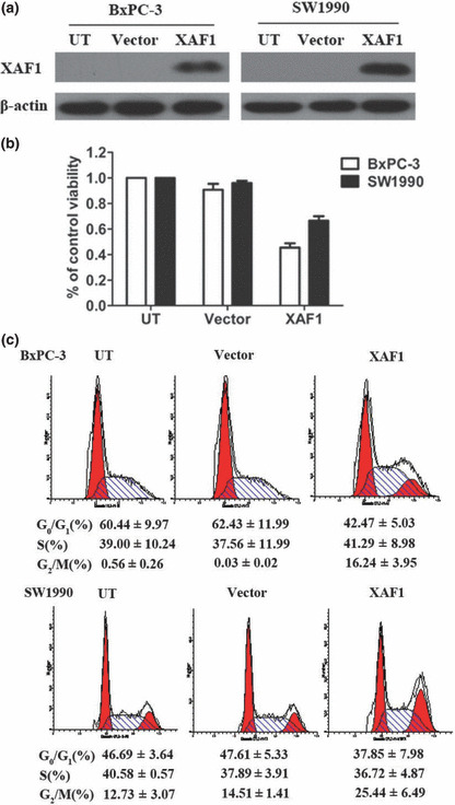
Effect of X chromosome‐linked inhibitor of apoptosis protein‐associated factor 1 (XAF1) overexpression on cell growth and cell cycle distribution of pancreatic cancer cell lines. (a) Western blot analysis showing expression of XAF1 in BxPC‐3 and SW1990 cells after they had been infected with an adenovirus containing XAF1 construct (XAF1). Untreated (UT) cells and cells infected with adenovirus alone (vector) served as the control. (b) Viability of BxPC‐3 and SW1990 cells evaluated by MTT assay in response to XAF1 overexpression. Columns, mean (n = 3); bars, SE. P < 0.05, significantly different compared with control. (c) Cell cycle distribution of BxPC‐3 and SW1990 cells infected with Ad5/F35‐XAF1 or vector. All assays were done three times, and in triplicate wells.