
Figure 4.
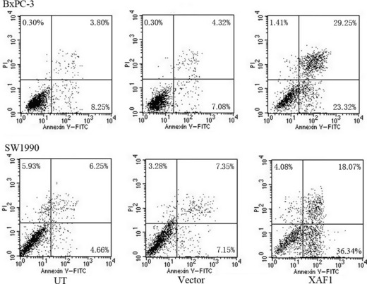
Analysis of apoptosis using flow cytometry in BxPC‐3 and SW1990 pancreatic cancer cells after treatment with an adenovirus containing X chromosome‐linked inhibitor of apoptosis protein‐associated factor 1 (XAF1) construct, Ad5/F35‐XAF1. Enhanced apoptosis was indicated by a shift to cells that were FITC+/propidium iodide (PI)−.