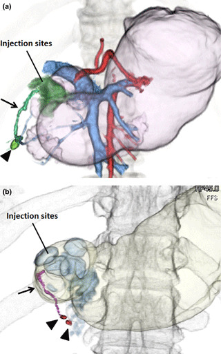
Figure 1.

Representative 3‐D computed tomography (CT) lymphograms for early gastric cancer. (a) A tumor was located on the lower side and posterior wall of the stomach (case number 1). The CT lymphography successfully detected an enhanced lymph node (arrowhead) and lymphatic flow toward the infrapyloric lymph node (arrow). Pictures of the portal and artery phases have been merged. (b) A tumor was located on the lower side and anterior wall of the stomach (case number 10). The CT lymphography successfully detected two enhanced lymph nodes (arrowheads) and lymphatics toward the infrapyloric lymph nodes (arrow).
