
Figure 2.
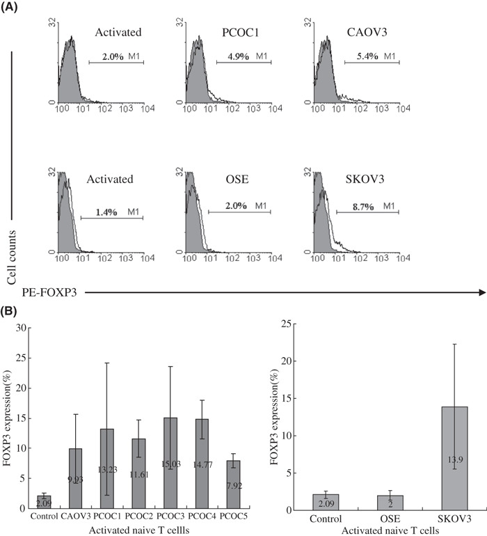
Supernatants from CAOV3 and ovarian carcinoma cell culture revealed the same function as that from SKOV3, but supernatant from ovarian surface epithelia cells (OSE)did not. (A) FOXP3 expression in activated naïve T cells treated with culture supernatant (CS) of PCOC1, CAOV3, or OSE on day 3 by flow cytometry. One representative staining is shown for at least three independent experiments. (B) The average percentage and range of FOXP3 expression in activated naïve T cells treated with CS of five PCOC, CAOV3, or OSE on day 3 from at least three independent experiments.