Figure 2.

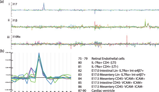
Expression of several interleukins (ILs) and interleukin receptors. (a) Probe‐pair signals for three interleukin‐related genes across a database containing 250 samples: (i) Il‐17, (ii) Il‐1β, and (iii) Il‐10rα. (b) A magnified view of the samples from which expression of Il‐17 can be seen. A clear signal can be seen in both samples 82 and 83, but in none of the other 248 samples in the database. The signal in sample 82 is around six‐times higher than that in 83. The strong deviation from the baseline, in combination with the high extent of correlation of individual probe‐pair signals, provide a high degree of confidence in the specificity of the expression. However, although there is about a six‐fold difference in the signal between 82 and 83, it is difficult to conclude that this represents the relative levels in the sample tissues, since we do not know by how much the signal tends to vary within these samples. In this specific case using replicate samples may not help much, as it is quite likely that the signal in 83 results from a cross‐contamination with tissue from 82, and the only way in which this can clearly be resolved is through in situ measurements.
