
Figure 1.
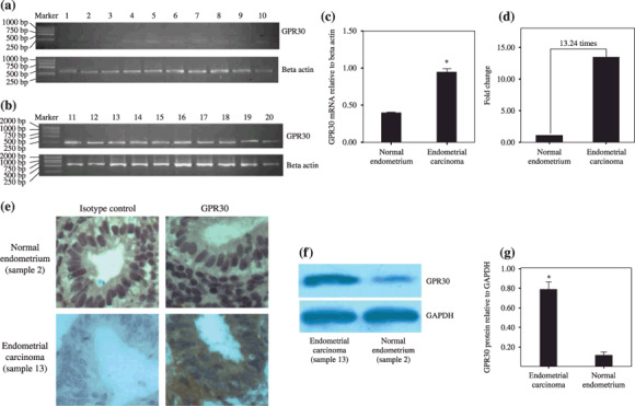
G protein‐coupled receptor (GPR) 30 transcription and overexpression in endometrial carcinoma. (a) Normal endometrium tissue: samples 1–6, endometrium of proliferative phase; samples 7–10, endometrium of secretory phase. (b) Endometrial carcinoma: samples 11–20, 10 carcinoma samples. Note: the marker contains the fragments (from top to foot of gel) 2000, 1000, 750, 500, 250, and 100 bp. (c) The y‐axis shows the ratio of optical density of GPR30 to β‐actin (mean ± SD) from (b). *P < 0.05. (d) The relative mRNA levels of GPR30 in normal endometrium and endometrial cancer by real‐time polymerase chain reaction. Ten normal endometrial tissues and 10 endometrial carcinoma tissues were used. (e) Immunochemistry for GPR30 in normal endometrium tissue and endometrial carcinoma. Normal endometrium, endometrial carcinoma, isotype control detected by rabbit IgG, and GPR30 expression detected by rabbit anti‐human GPR30 antibody are shown. Magnification, ×400. (f) Western blotting analysis for GPR30 protein levels in normal endometrium tissue and endometrial carcinoma. (g) showed the densitometric analysis. *P < 0.05. Fifty endometrial carcinoma samples and 17 normal endometrium samples were used in the reverse transcription–polymerase chain reaction, immunochemistry, and western blotting. The results were highly reproducible and these pictures are representative. GAPDH, glyceraldehyde‐3‐phosphate dehydrogenase.