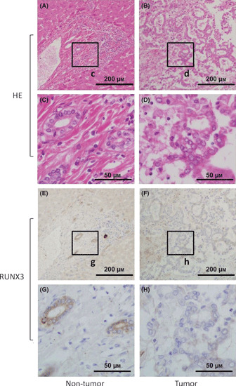
Figure 1.

(A–D) Representative hematoxylin and eosin (HE) and (E–H) immunohistochemical stainings of RUNX3 in biliary tract cancer specimen, including non‐tumor tissue, are shown. The c, d, g, h boxes indicate the positions of the enlargements shown below. Scale bars in (A,B,E,F) panels: 200 μm; (C,D,G,H) panels: 50 μm.
