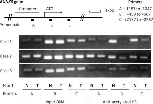
Figure 2.

The top panel is a schematic presentation of the RUNX3 gene, indicating the location of three primer pairs used for PCR amplification in ChIP assays. Chromatin fragments were immunoprecipitated with an antibody against acetylated histone H3 in biliary tract cancer specimens. Input DNA, without immunoprecipitation by anti‐acetylated H3 antibody, was also subjected to PCR as a positive control. The right part of the bottom three panels shows representative ChIP assays prepared using anti‐deacetylated H3. Cases 1 and 2 show lower accumulations of acetylated histone H3 in chromatin associated with RUNX3 in tumors, especially in primer B. N, non‐tumor; T, Tumor.
