Figure 3.

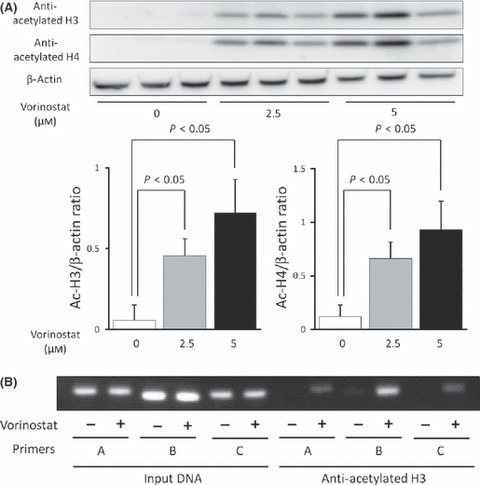
(A) Vorinostat stimulated histone acetylation in a dose‐dependent manner. Mz‐Ch‐A2 cells were treated with 0–5 μM vorinostat for 48 h. Cells were harvested and histones were prepared as described. Histone acetylation was detected by Western blot with antibodies against acetylated H3 and H4. Densitometric ratios of H3 and H4 are also shown. *P < 0.05 when compared with the value derived by non‐stimulation. (B) Vorinostat‐induced accumulation of acetylated histone H3 in chromatin associated with RUNX3. Three primer pairs described in Fig. 2 were used for PCR amplification in ChIP assays. Chromatin fragments from cells cultured with or without 5 μM vorinostat for 48 h were immunoprecipitated with an antibody against acetylated histone H3 in Mz‐Ch‐A2 cells. The panel shows ChIP assays prepared using anti‐deacetylated H3. Input DNA, without immunoprecipitation by anti‐acetylated H3 antibody, was also subjected to PCR as a positive control.
