
Figure 5.
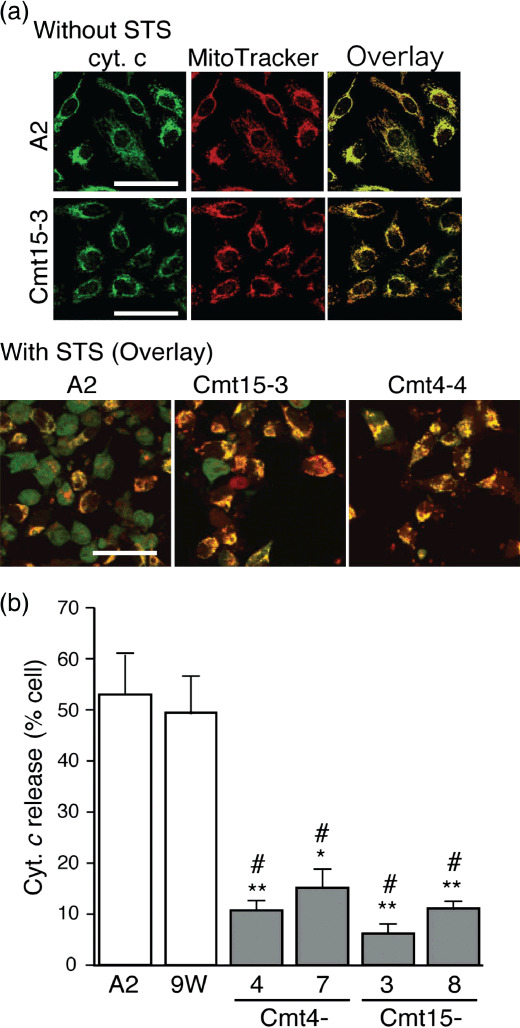
Mutant mitochondrial DNAs (mtDNAs) inhibited cytochrome (cyt.) c release. Cells were incubated in complete medium with or without staurosporine (STS) (0.5 µM) for 4 h and then treated with MitoTracker Red (red). After fixing, permeabilizing and blocking, the cells were treated with anti‐cyt. c mouse monoclonal antibody (250‐dilution; Promega, Madison, WI, USA), followed by incubation with antimouse IgG BODIPY (green) as a secondary antibody as described.(24) The cells were imaged with a confocal laser scanning microscope. (a) Upper panels: representative images of cells without STS treatment at high magnification. Lower panels: representative overlaid images of cells with STS treatment. Cells exhibiting cyt. c release were diffusely stained green. In contrast, cells without cyt. c release were stained yellow (overlay of green and red) Bar: 50 µm. (b) Population of cells exhibiting cyt. c release. Approximately 300 cells per experiment were examined to obtain the ratio of cells exhibiting cyt. c release to the total cell number. Means of the ratio from three independent experiments are shown with SD (vertical bars). Bar: 100 µm. *P < 0.05, **P < 0.001, versus A2; #P < 0.001, versus 9 W.