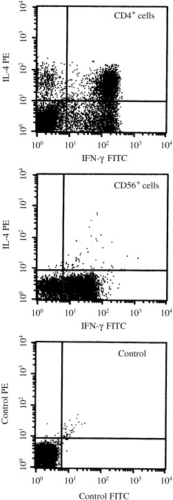
Figure 5.

Intracytoplasmic cytokine staining of CD4+ and CD56+ cells. PBMC from case 1 were stimulated with phorbol myristate acetate and Ca ionophore. The cells were triple stained with anti‐IFN‐γ, anti‐IL‐4, and anti‐CD4 or anti‐CD56. CD4+ or CD56+ cells were gated so that cytokine production could be seen in each cell population. The control was carried out with isotype‐matched monoclonal antibodies labeled with FITC and PE.
