
Figure 4.
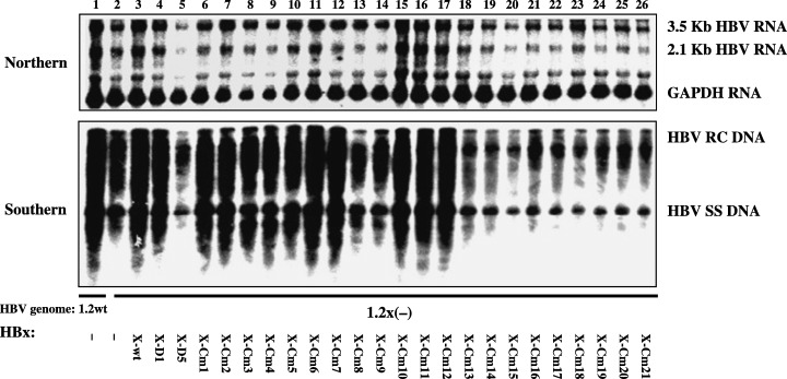
Mapping of the HBx sequences required for the stimulation effect on HBV transcription and replication. HepG2 cells were transiently transfected with the wild‐type HBV construct payw1.2 (1.2wt, lane 1) or the HBx‐minus HBV construct payw (42) 7 (1.2X(–), lanes 2–26) plus empty vector control (–, lanes 1 and 2), full‐length HBx expression vector (X‐wt, lane 3), different truncated HBx expression vectors (X‐D1, lane 4; X‐D5, lane 5), or a series of clustered mutated HBx expression vectors (X‐Cm1 to X‐Cm21, lanes 6–26, respectively). Top is RNA (Northern) hybridization analysis of HBV transcripts. The glyceraldehyde 3‐phosphate dehydrogenase (GAPDH) transcript was used as an internal control for RNA loading per lane. Bottom is DNA (Southern) hybridization analysis of HBV replication intermediates. HBV RC DNA, HBV relaxed circular DNA; HBV SS DNA, HBV single‐stranded DNA. The results were according to the report by Tang et al.( 27 )