
Figure 6.
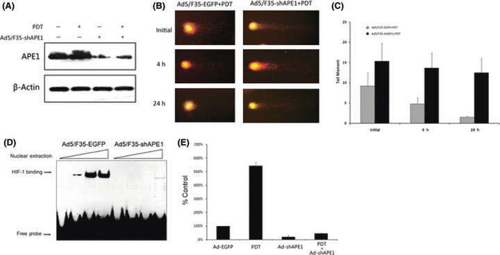
Both DNA repair and redox activities were involved in cellular survival after photodynamic irradiation. The Ad5/F35‐shAPE1‐infected or Ad5/F35‐EGFP‐infected groups received 4.5 kJ/m2 photodynamic irradiation at 48 h after infection. Then, APE1 protein level was assayed by western blotting at 12 h after irradiation. (A) The upregulation of APE1 induced by photodynamic therapy (PDT) was significantly abolished after Ad5/F35‐shAPE1 infection. (B) DNA single‐strand break at initial (immediately after irradiation), 4, and 24 h after PDT in the Ad5/F35‐shAPE1 and control groups using alkaline comet assay. (C) The ‘tail’ of the comet, which was quantified by Komet software, was considered as the impaired DNA fragment. It demonstrated that the repair capacity to this type of DNA lesion was significantly suppressed by Ad5/F35‐shAPE1 infection and the DNA damage caused by PDT was not completely repaired at 24 h post‐irradiation, which was repaired at 4 h in the control group. We then assayed the transcription factor redox regulation activity of APE1 by (D) EMSA and (E) RT‐PCR 12 h after PDT. Oligonucleotide containing HIF‐1 consensus was used to assay the DNA binding activity of HIF‐1, which is regulated by APE1. It was shown that the DNA binding activity was significantly elevated after PDT and inhibited by Ad5/F35‐shAPE1 infection. The expression of VEGF, an important downstream gene of HIF‐1, was then assayed by real‐time RT‐PCR. In accordance with EMSA results, real‐time RT‐PCR indicated that the expression of VEGF was significantly upregulated by PDT and downregulated by Ad5/F35‐shAPE1. Results were obtained from three independent experiments.