
Figure 5.
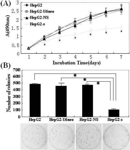
Cell proliferation determined by 3‐(4,5‐dimethyl‐2‐thiazolyl)‐2, 5‐diphenyl‐2H‐tetrazolium bromide (MTT) and colony formation assays. (A) The protracted cell growth curve and the results of the inhibitory rates of cell growth were applied to absorbance at 490 nm. The proliferation of HepG2‐s cells was significantly suppressed in a time‐dependent manner, and the highest inhibition rate was 40.2 ± 2.5% (*P < 0.01) on day 7. (B) The HepG2‐s cells showed much less colonies than HepG2‐CMVneo, HepG2‐NS and untransfected HepG2 cells. These experiments were repeated three times, *P < 0.05.