
Figure 2.
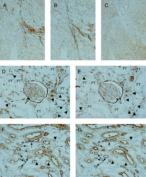
Immunohistochemistry for the three antigens on the serial sections of cancerous and normal renal tissues. (A–C) Serial sections of G2 renal cell carcinoma (original magnification ×14). Positive staining for S100A10 (A) and annexin II (B) is diffusely found on the plasma membrane and occasionally appeared faintly in the cytoplasm. Immunopositivity of B‐FABP (C) is seen in the cytoplasm of the carcinoma cells. (D, E) Normal renal cortex (original magnification ×14). Bowman's capsule and some of the distal convoluted tubules (asterisks) and glomerular podocytes (arrows) are positively stained for S100A10 (D) and annexin II (E). Immunostaining is also found in endothelial cells (arrowheads). The few proximal tubules (PT) represent the signals. (F, G) Normal renal medulla (original magnification ×14). The immunopositivity for S100A10 (F) is in agreement with that for annexin II (G). Positive reactions observed are mainly associated with the plasma membrane of the collecting duct (asterisks) and the thin portion of Henle's loop (arrows). Endothelium (arrowheads) is also diffusely positive for both proteins.