
Figure 1.
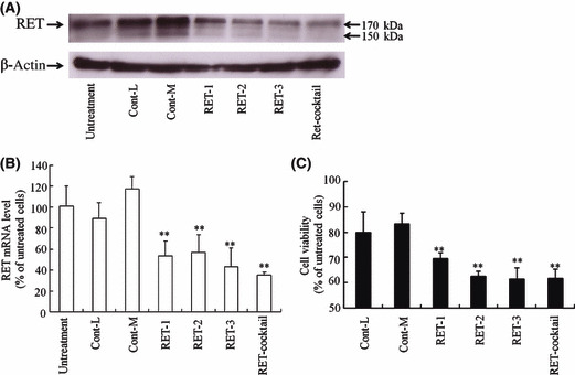
Inhibition of RET expression by transfection of RET siRNA with Lipofectamine 2000 into TT cells. Expressions of RET protein (170 KDa) and mRNA were detected by Western blot (A) and quantitative RT‐PCR (B) analyses, respectively, 72 h after transfection with 100 nm Cont‐L and ‐M siRNA, RET‐1, ‐2 and ‐3 siRNA and their cocktail. The number of viable cells was determined by WST‐8 assay 72 h after transfection (C). n = 4 for each sample. **P < 0.01, compared with Cont‐L siRNA transfected cells.