
Figure 5.
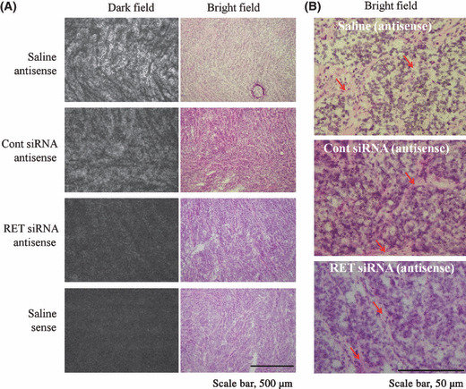
In situ hybridization of a 35S‐labeled antisense or sense probe generated against RET mRNA. An antisense probe was used to detect RET mRNA. The sense probe for the RET gene was used as a negative control probe. In (A), silver grains in a dark field indicate RET mRNA expression. Scale bar: 500 μm. In (B), microautoradiographs in bright fields in Fig. 5(A) were enlarged. Black grains indicate RET mRNA expression. Red arrows indicate stroma cells. Scale bar: 50 μm.