
Figure 1.
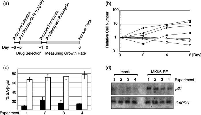
MKK6‐expressing WI‐38 cells used for microarray analyses. (a) Experimental schemes showing sample preparation for microarray. Normal human fibroblast WI‐38 cells were infected with RasV12‐expressing retrovirus or empty vector. Infected cells were selected by puromycin treatment and harvested as indicated. (b) Growth curves of MKK6‐EE‐expressing WI‐38 cells and empty‐vector‐infected cells. Retroviral infections were done four times (represented by triangles, circles, diamonds and squares). Open and filled symbols indicate MKK6‐EE‐expressing and mock‐treated cells, respectively. (c) Percentages of SA‐β‐gal‐positive cells at day 6. White and black bars represent MKK6‐EE‐expressing and mock‐treated cells, respectively. (d) Expression levels of CDK inhibitor p21 and GAPDH in MKK6‐EE‐expressing and mock‐treated cells as revealed by northern blotting analysis.